Noonan Syndrome
What is Noonan Syndrome?
Noonan syndrome is a disorder that involves unusual facial characteristics, short stature, heart defects present at birth, bleeding problems, developmental delays, and malformations of the bones of the rib cage.
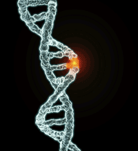
Noonan syndrome is caused by changes in one of several autosomal dominant genes. A person who has Noonan syndrome may have inherited an altered (mutated) gene from one of his or her parents, or the gene change may be a new change due to an error carried by the egg or sperm or occurring at conception. Alterations in four genes – PTPN11, SOS1, RAF1 and KRAS – have been identified to date.
Noonan syndrome is present in about 1 in 1,000 to 1 in 2,500 people.
What are the symptoms of Noonan Syndrome?
Symptoms of Noonan syndrome may include the following:
- A characteristic facial appearance.
- Short stature
- Heart defect present at birth (congenital heart defect)
- A broad or webbed neck
- Minor eye problems such as strabismus in up to 95 percent of individuals
- Bleeding problems such as a history of abnormal bleeding or bruising
- An unusual chest shape with widely-spaced and low set nipples
- Developmental delay of varying degrees, but usually mild
- In males, undescended testes (cryptorchidism)
How is Noonan syndrome diagnosed?
The diagnosis of Noonan syndrome is based on the person’s clinical symptoms and signs. The specialist examines the person looking for the specific features of Noonan syndrome.
Individuals who have Noonan syndrome have normal chromosome studies. Four genes – PTPN11, SOS1, RADF1 and KRAS – are the only genes that are known to be associated with Noonan syndrome. Approximately 50 percent of individuals with Noonan syndrome have mutations in the PTPN11 gene. Twenty percent of those with Noonan Syndrome have mutations in the SOS1. Mutations in the RAF1 gene account for between 10 and 15 percent of Noonan syndrome cases. About 5 percent of people with Noonan syndrome have mutations in the KRAS gene and usually have a more severe or atypical form of the disorder. The cause of Noonan syndrome in the remaining 10 to 15 percent of people with this disorder is not yet known.
What is the treatment for Noonan syndrome?
Treatment for individuals who have Noonan syndrome is based on their particular symptoms. Heart problems are treated in the same way as they are for individuals in the general population. Early intervention programs are used to help with developmental disabilities, when present. Bleeding problems that can be present in Noonan syndrome may have a variety of causes and are treated according to their cause. Growth problems may be caused by lack of growth hormone and may be treated with growth hormone treatment. Symptoms such as heart problems are followed on a regular basis.
Is Noonan syndrome inherited?
Noonan syndrome is inherited in families in an autosomal dominant pattern. This means that a person who has Noonan syndrome has one copy of an altered gene that causes the disorder. In about one-third to two-thirds of families one of the parents also has Noonan syndrome. The parent who has Noonan syndrome has a 1 in 2 (50 percent) chance to pass on the altered gene to a child who will be affected; and a 1 in 2 (50 percent) chance to pass on the normal version of the gene to a child who will not have Noonan syndrome. In many individuals who have Noonan syndrome, the altered gene happens for the first time in them, and neither of the parents has Noonan syndrome. This is called a de novo mutation. The chance for these parents to have another child with Noonan syndrome is very small (less than 1 percent).
Thalassemia
What do we know about heredity and thalassemia?
Wilson Thalassemia is actually a group of inherited diseases of the blood that affect a person’s ability to produce hemoglobin, resulting in anemia. Hemoglobin is a protein in red blood cells that carries oxygen and nutrients to cells in the body. About 100,000 babies worldwide are born with severe forms of thalassemia each year. Thalassemia occurs most frequently in people of Italian, Greek, Middle Eastern, Southern Asian and African Ancestry.
The two main types of thalassemia are called “alpha” and “beta,” depending on which part of an oxygen-carrying protein in the red blood cells is lacking. Both types of thalassemia are inherited in the same manner. The disease is passed to children by parents who carry the mutated thalassemia gene. A child who inherits one mutated gene is a carrier, which is sometimes called “thalassemia trait.” Most carriers lead completely normal, healthy lives.
A child who inherits two thalassemia trait genes – one from each parent – will have the disease. A child of two carriers has a 25 percent chance of receiving two trait genes and developing the disease, and a 50 percent chance of being a thalassemia trait carrier.
Most individuals with alpha thalassemia have milder forms of the disease, with varying degrees of anemia. The most severe form of alpha thalassemia, which affects mainly individuals of Southeast Asian, Chinese and Filipino ancestry, results in fetal or newborn death.
A child who inherits two copies of the mutated gene for beta thalassemia will have beta thalassemia disease. The child can have a mild form of the disease, known as thalassemia intermedia, which causes milder anemia that rarely requires transfusions.
Thalassemia Major: A Serious Disorder
The more severe form of the disease is thalassemia major, also called Cooley’s Anemia. It is a serious disease that requires regular blood transfusions and extensive medical care.
Those with thalassemia major usually show symptoms within the first two years of life. They become pale and listless and have poor appetites. They grow slowly and often develop jaundice. Without treatment, the spleen, liver and heart soon become greatly enlarged. Bones become thin and brittle. Heart failure and infection are the leading causes of death among children with untreated thalassemia major.
The use of frequent blood transfusions and antibiotics has improved the outlook for children with thalassemia major. Frequent transfusions keep their hemoglobin levels near normal and prevent many of the complications of the disease. But repeated blood transfusions lead to iron overload – a buildup of iron in the body – that can damage the heart, liver and other organs. Drugs known as “iron chelators” can help rid the body of excess iron, preventing or delaying problems related to iron overload.
Thalassemia has been cured using bone marrow transplants. However, this treatment is possible only for a small minority of patients who have a suitable bone marrow donor. The transplant procedure itself is still risky and can result in death.
Gene Therapy Offers Hope for a Cure
Scientists are working to develop a gene therapy that may offer a cure for thalassemia. Such a treatment might involve inserting a normal beta globin gene (the gene that is abnormal in this disease) into the patient’s stem cells, the immature bone marrow cells that are the precursors of all other cells in the blood.
Another form of gene therapy could involve using drugs or other methods to reactivate the patient’s genes that produce fetal hemoglobin – the form of hemoglobin found in fetuses and newborns. Scientists hope that spurring production of fetal hemoglobin will compensate for the patient’s deficiency of adult hemoglobin.
Is there a test for thalassemia?
Blood tests and family genetic studies can show whether an individual has thalassemia or is a carrier. If both parents are carriers, they may want to consult with a genetic counselor for help in deciding whether to conceive or whether to have a fetus tested for thalassemia.
Prenatal testing can be done around the 11th week of pregnancy using chorionic villi sampling (CVS). This involves removing a tiny piece of the placenta. Or, the fetus can be tested with amniocentesis around the 16th week of pregnancy. In this procedure, a needle is used to take a sample of the fluid surrounding the baby for testing.
Assisted reproductive therapy is also an option for carriers who don’t want to risk giving birth to a child with thalassemia. A new technique, pre-implantation genetic diagnosis (PGD), used in conjunction with in vitro fertilization, may enable parents who have thalassemia or carry the trait to give birth to healthy babies. Embryos created in-vitro are tested for the thalassemia gene before being implanted into the mother, allowing only healthy embryos to be selected.
Download this Issue
To Download a PDF file version of this Issue of the NASET’sGenetics in Special Education Series – CLICK HERE

