Neurofibromatosis
What is neurofibromatosis?
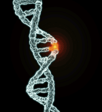
Neurofibromatosis (NF) is a genetic neurological disorder that can affect the brain, spinal cord, nerves and skin. Tumors, or neurofibromas, grow along the body’s nerves or on or underneath the skin. Scientists have classified NF into two distinct types: neurofibromatosis type 1 (NF1) and NF2. NF1, formerly known as von Recklinghausen’s NF, is the more common of the types. It occurs in approximately 1 in 4,000 births. NF2, also referred to as bilateral acoustic NF, central NF or vestibular NF, occurs less frequently- 1 in 40,000 births. Occurences of NF1 and NF2 are present among all racial groups and affect both sexes equally. The tumors arise from changes in the nerve cells and skin cells. Tumors also may press on the body’s vital areas as their size increases. NF may lead to developmental abnormalities and/or increased chances of having learning disabilities. Other forms of NF, where the symptoms are not consistent with that of NF1 or NF2, have been observed. A rare form of NF is schwannomatosis. However, the genetic cause of this form of NF has not been found.
What are the symptoms of neurofibromatosis?
Symptoms for neurofibromatosis type 1 include:
- Presence of light brown sports (café-au-lait) on the skin.
- Appearance of two or more neurofibromas (pea-sized bumps) that can grow either on the nerve tissue, under the skin or on many nerve tissues.
- Manifestation of freckles under the armpits or in the groin areas.
- Appearance of tiny tan clumps of pigment in the iris of the eyes (Lisch nodules).
- Tumors along the optic nerve of the eye (optic glioma).
- Severe curvature of the spine (scoliosis).
- Enlargement or malformation of other bones in the skeletal system.
Symptoms for NF1 vary for each individual. Those that are skin-related are often present at birth, during infancy and by a child’s tenth birthday. From ages 10 to 15, neurofibromas may become apparent. Symptoms such as café-au-lait spots, freckling and Lisch nodules pose minimal or no health risk to a person. Though neurofibromas are generally a cosmetic concern for those with NF1, they can sometimes be psychologically distressing. For 15 percent of individuals with NF1, the symptoms can be severely debilitating. Neurofibromas can grow inside the body and may affect organ systems. Hormonal changes at puberty and/or even pregnancy may increase the size of neurofibromas. Nearly 50 percent of children with NF1 have speech problems, learning disabilities, seizures and hyperactivity. Less than one percent of those affected with NF1 may have malignant tumors and may require treatment.
Symptoms for neurofibromatosis type 2 include:
- Tumors along the eighth cranial nerve (schwannomas).
- Meningiomas and other brain tumors.
- Ringing noises inside the ear (tinnitus), hearing loss and/or deafness.
- Cataracts at a young age.
- Spinal tumors.
- Balance problems.
- Wasting of muscles (atrophy).
Individuals with NF2 develop tumors that grow on the eighth cranial nerves and on the vestibular nerves. These tumors often cause pressure on the acoustic nerves, which result in hearing loss. Hearing loss may begin as early as an individual’s teenage years. Tinnitus, dizziness, facial numbness, balance problems and chronic headaches may also surface during the teenage years. Numbness may also occur in other parts of the body, due to spinal cord tumors.
The rare form of NF, schwannomatosis, which was recently identified, does not develop on the eighth cranial nerves, and does not cause hearing loss. It causes pain primarily, and in any part of the body. Though schwannomatosis may also lead to numbness, weakness or balance problems like NF1 or NF2, the symptoms are less severe.
How is neurofibromatosis diagnosed?
Neurofibromatosis is diagnosed from a combination of findings. For children to be diagnosed with NF1, they must show at least two of the aforementioned symptoms associated with NF1. A physical examination by a doctor familiar with the disorder is usually performed. Doctors may use special lamps to examine the skin for café-au-lait spots. Doctors may also rely on magnetic resonance imaging (MRI), X-rays, computerized tomography (CT scan) and blood tests to detect defects in the NF1 gene.
For NF2, doctors will pay close attention to hearing loss. Hearing tests as well as imaging tests are used to look for tumors in and around the auditory nerves, the spinal cord or the brain. Audiometry and brainstem auditory evoked response tests can help determine whether the eighth cranial nerve is functioning properly. Family history of NF2 is also a key focal area for diagnosis.
Genetic testing is also used to diagnose NF1 and NF2. Testing conducted before birth (prenatal) is helpful to identify individuals who have a family history of the disorder, but do not yet have the symptoms. Still, gene tests have no way of predicting the severity of NF1 or NF2. Genetic testing is performed by either direct gene mutation analysis and/or linkage analysis. Mutation analysis looks to identify the particular gene changes that cause NF. A linkage analysis is useful if the mutation analysis does not provide enough conclusive information. With at linkage analysis, blood tests from multiple family members are taken to track the chromosome that carry the disease-causing gene through two or more generations. Linkage testing is around 90 percent accurate in determining whether individuals have NF. Mutation analysis is 95 percent accurate in finding a mutation for NF1, and 65 percent accurate for NF2.
How is neurofibromatosis treated?
Though there is no cure for either NF1 or NF2, there are ways to treat the effects the disease. Surgery may be helpful in removing tumors, though there is a risk of the tumors regenerating. For optic gliomas, treatment may include surgery and/or radiation. For scoliosis, treatment may include surgery or back braces. For symptoms associated with NF2, surgery may be a viable option, however not without complications that could result in additional loss of hearing or deafness. Hearing aids are ineffective when parts of the auditory nerve are removed. A breakthrough in treatment became available recently to NF2 patients, when the Food and Drug Administration approved an Auditory Brainstem Implant [fda.gov] for those who have parts of their auditory nerve removed and have suffered from subsequent hearing loss. The implant transmits sound signals to the brain directly and allows people to hear certain sounds and speech . Radiation treatment, may also help relieve symptoms associated with NF2.
What do we know about heredity and Neurofibromatosis?
Neurofibromatosis can either be an inherited disorder or the product of a gene mutation. Both NF1 and NF2 are caused by two separate abnormal genes and may be inherited from parents who have NF or may be the result of a mutation in the sperm or egg cells. NF is considered an autosomal dominant disorder because the gene is located on one of the 22 chromosome pairs, called autosomes.The gene for NF1 is located on chromosome 17. The gene for NF2 is located on chromosome 22. Children have a 50 percent chance of inheriting the genes that cause NF if the parent has NF. The type of NF the child inherits will be the same as that of the parent. Therefore, if the parent has NF1, there will be a 50 percent chance the child will have NF1. If the parent has NF2, there will be a 50 percent chance the child will have NF2. The only difference between the child and the parent in these circumstances is the severity of NF and the appearance of symptoms. The presence of only one changed or affected gene can cause the disorder to appear. However, the action of the unaffected gene that is paired with the dominant gene does not prvent the disorder from appearing. People with NF can make two different kinds of reproductive cells: one that can cause a child to have NF and the other that will produce an unaffected child, if that is the gene that happens to be used. When an unaffected individual conceives a child with a person with NF, there are four possible cell combinations – two combinations that will yield a child with NF and the other two that will yield an unaffected child.
Holoprosencephaly
What do we know about holoprosencephaly?
Holoprosencephaly (HPE) is a relatively common birth defect of the brain, which often can also affect facial features, including closely spaced eyes, small head size, and sometimes clefts of the lip and roof of the mouth, as well as other birth defects. Holoprosencephaly is a disorder caused by the failure of the prosencephalon (the embryonic forebrain) to sufficiently divide into the double lobes of the cerebral hemispheres. The result is a single-lobed brain structure and severe skull and facial defects. In most cases of holoprosencephaly, the malformations are so severe that babies die before birth. In less severe cases, babies are born with normal or near-normal brain development and facial deformities that may affect the eyes, nose and upper lip.
This birth defect occurs soon after conception. It has a prevelance of 1 in 250 during early embryo development, and 1 in 10,000 to 1 in 20,000 at term.
There are three classifications of holoprosencephaly:
- Alobar, in which the brain has not divided at all, is usually associated with severe facial features.
- Semilobar, in which the brain’s hemispheres have somewhat divided, causes an intermediate form of the disorder.
- Lobar, in which there is considerable evidence of separate brain hemispheres, is the least severe form. In some cases of lobar holoprosencephaly the baby’s brain may be nearly normal.
The milder craniofacial characteristics of HPE include microcephaly, midface flattening, hypotelorism (closely spaced eyes), flat nasal bridge, and single maxillary central incisor. Approximately 80 percent of severe HPE have characteristic facial features. The least severe of the facial anomalies in holoprosencephaly is the median cleft lip (premaxillary agenesis). The most severe is cyclopia, an abnormality characterized by a single eye located in the area normally occupied by the root of the nose, and a missing nose or a proboscis (a tubular-shaped nose) located above the eye. The least common facial anomaly is ethmocephaly, in which a proboscis separates closely-set eyes. Cebocephaly, another facial anomaly, is characterized by a small, flattened nose with a single nostril situated below incomplete or underdeveloped closely-set eyes.
Not all individuals with HPE are affected to the same degree, even in families where more than one individual has this predisposition. This is why it is often helpful to discuss these issues with a professional in genetics who is trained to recognize features that might suggest that HPE is, or is not, likely to occur again in a family. The risk of reoccurrence is small in most families. There are a number of causes of HPE, including genetic alterations and environmental effects. The cause of HPE in any individual family is often unknown.
Is there a test for holoprosencephaly?
The best diagnostic procedure is a brain scan (CT or MRI). Molecular testing for several HPE genes are available.
Is there a treatment for holoprosencephaly?
Each child has a unique degree of malformations. Treatment must be individualized, although common problems occur. In general, treatment is largely symptomatic and supportive. Involvement in support groups and HPE Conferences are helpful (See: Additional Resources).
What is the prognosis?
The prognosis for individuals with the disorder depends on the severity of the brain and facial malformations and associated clinical complications. The older literature suggested that the prognosis was uniformly poor. Recent studies show a broader range of outcomes than previously assumed.
Download this Issue
To Download a PDF file version of this Issue of the NASET’sGenetics in Special Education Series – CLICK HERE